search
ouroboros-arbor

Motion of the Vertebrae in the Traditional Anatomical Planes


By Janis Savlovskis, MD

The question "How exactly moves each separate vertebra within the spine" may seem simple, but the answer is never trivial and requires mental flexibility. The reason for that is evident: the spine movements result from the sum of the motion of each separate vertebra relative to each other with the six degrees of freedom and dynamic instantaneous axis of rotation.

Six degrees of freedom passing through the functional spinal unit L3–L4
The image demonstrates the types of displacement (rotations and translations) of the upper vertebra relative to the lower vertebra in the standard anatomical three-dimensional coordinate system. Please note that the axes of motions never collide in a single point of space in real life. An exact position of the rotational axis may change during different phases of the same motion, i.e., the instantaneous axis of rotation has dynamic spatial coordinates that are affected by both – the type and the direction of movement.

Many approaches are used to answer the question about the range of motion of separate vertebrae. They could be summarized in the following three groups:

  • 1) the ex vivo studies that rely on the human spine specimens usually dissected and isolated from the rest of the body.
  • (2) the in vivo approachesthat measures the motions of the vertebrae by the use of non-invasive (monoplanar, biplanar x-ray, computed tomography, magnetic resonance, or combination of these methods) or invasive techniques (by inserting the fine metallic pins into the vertebrae and by tracking them during the motion).
  • (3) the in vivo indirect approach – the measurement of back curvature in different body positions and interpolating these results to vertebral motion.

Each method has some flaws that are hard to overcome:

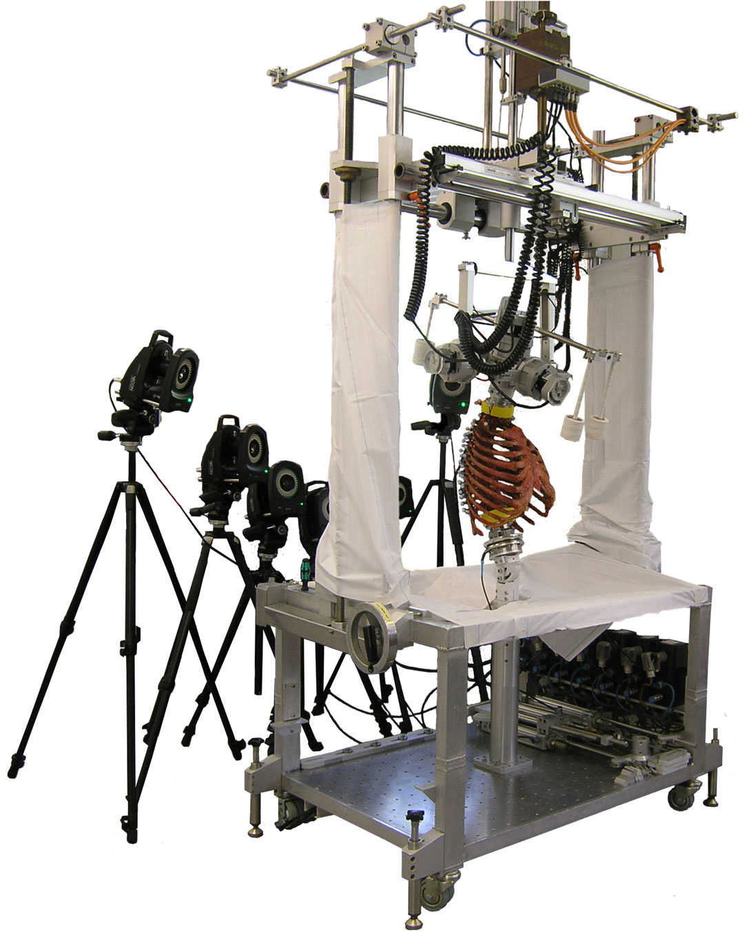
Ex vivo studies are precise (in terms of the measurement error). Still, they cannot fully simulate the physiological conditions irreversibly affected by the specimen preparation process (surrounding bone and soft tissue removal) and inevitable post mortem change of the soft tissue physical properties. Another limitation is the advanced age of subjects being included in the study. It is well established that the average range of the spine motion gradually reduces after the 4th decade of life (Dreischarf 2014; Arshad 2018) and don't match exactly the ROM of young individuals.

Human thorax in the experimental setup. Image from the article
An experimental setup for the ex vivo measurement of the spine motions, using an optical motion tracking system with six cameras. (Image from the open-access article by Liebsch et al, 2017)

The studies based on the medical imaging techniques (x-Ray, Computed Tomography) are not entirely safe due to the ionizing radiation or are too expensive (Magnetic Resonance) to recruit a large number of healthy volunteers. Another issue is that both – Computed Tomography and Magnetic Resonance – are done in a supine position within the limited space. The investigated spine positions are relatively passive (relaxed musculature), and may not match precisely the active and vertically balanced movements observed in routine activities.

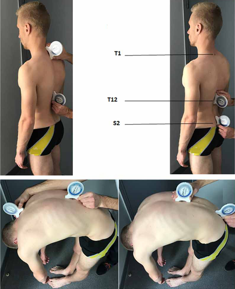
Finally, the externally applicable devices (inclinometers, goniometers, accelerometers, etc.) for the estimation of spine curvature cannot directly track individual vertebrae. The measurements performed in these studies are usually segmental, and their results rely heavily on the interpolation. For example, measuring the dynamic curvature of upper vs. mid- vs. lower thoracic spine then is interpolated to the motion of Th1–Th3 vs. Th4–Th7 vs. Th8–Th12.

Use of inclinometers. Image from the article
Positioning of the two inclinometers for the evaluation of the spine range of motion (Image from the open-access article by Ditcharles et al, 2017)

The meta-analysis presented here includes all types of studies.

The demonstrated images show the absolute difference in an individual vertebra's spatial position relative to the neutral spine. When these small respective motions sum-up, we see the resulting movements of the spine as a whole.

The graphs are built following the uniform logic:

An example of graph with comments

The reported results were normalized by converting the angular scale to the fractional (percentage) scale to reduce the variance. Studies are usually focused on the examination of the single segment of the spine – atlanto-occipital (C0–C1), subaxial (C2–C7), thoracic (Th1–Th11), or lumbar segment (L1–L5). To preserve the comparability of the ROM implemented in the Anatomy Standard spine model with the literature data, the reported percentage values were calculated to reflect the fraction of vertebral motion relative to the corresponding part of the spine, and not relative to the entire spine. Due to methodological limitations, the mobility of the last thoracic vertebra remains unreported. To avoid the distortion bias, the fraction of motion for this vertebra was calculated proportionally and out of the thoracic spine limit. All the graphs and images presented on this page are designed to show both – original and normalized data. Click over to switch between the angle scale and fractional (percentage) scale.

Motioin of Vertebrae in the Sagittal Plane (Flexion & Extension)


Flexion and Extension of Entire Spine


Icon of crossfade image
The flexion and extension range of motion for all vertebra Graph, demonstrating the evidence for the flexion-extension ROM of all vertebra
The lateral aspect of vertebrae demonstrating the combined flexion / extension range of motion of vertebrae relative to the neutral spine. The sagittal inclination angle of vertebrae in neutral spine is preserved
An absolute and relative range of motion from the full flexion to the full extension. Click an image to switch between an absolute (°) and relative (%) scale

The evidence for range of combined flexion / extension motion of thoracic vertebrae is very low. There are only three literature sources available, and the results of these studies are mutually highly controversial. By the contrast – there are multiple in-vivo studies for the thoracic spine flexion and extension done using inclinometer or goniometer. The results of these studies are revieled in the following chapter ↴

Flexion and Extension of the Thoracic Vertebrae


This section clarify the motion of the thoracic vertebrae by analysing separately flexion and extension of the thorax.

Icon of crossfade image
Flexion and extension ROM of the thoracic spine Graphs demonstrating the basis for the angular dislocation of the thoracic vertebrae
The lateral aspect of the thoracic vertebrae demonstrating separately the range of flexion and the range of extension relative to their position in the neutral spine. The sagittal inclination angle of vertebrae in neutral spine is preserved
An absolute and relative flexion and flexion of the thoracic vertebrae. Click the graph to switch between an absolute (°) and relative (%) scale

Motioin of Vertebrae in the Frontal Plane (Lateral Bending)


Icon of crossfade image
Lateral bending ROM for all the vertebrae of the spine Graph, demonstrating the lateral bending for all the vertebrae
The anterior aspect of the vertebrae, demonstrating their absolute lateral bending range. The sagittal inclination angle of vertebrae in neutral spine is preserved for both positions (neutral and laterally flexed)
An absolute and relative lateral flexion of vertebrae implimented in the Anatomy Standard spine model in context with the scientific literature data. (Click the graph to switch between an absolute (°) and relative (%) scale)

Motioin of Vertebrae in the Horizontal Plane (Axial Rotation)


Icon of crossfade image
Axial rotation of individual veretbrae within the spine Graph demonstrating the scientifica basis for the axial rotation of vertebrae
The antero-superior view to the spine demonstrating an absolute axial rotation of each vertabra. The sagittal inclination angle of vertebrae seen in neutral spine is preserved for both positions (neutral and axially rotated).
An absolute and relative axial rotation of vertebrae implimented in the Anatomy Standard spine model in context with the scientific literature data. (Click the graph to switch between an absolute (°) and relative (%) scale)

List of references

  • Aho A, et al. Segmentary mobility of the lumbar spine in antero-posterior flexion. Ann Med Intern Fennieae, 1955, 44(4):275–285.
  • Alqhtani R, et al. Reliability of an accelerometer-based system for quantifying multiregional spinal range of motion. Journal of Manipulative and Physiological Therapeutics. 2015, 38(4):275-281.
  • Amevo B, et al. Instantaneous axes of rotation of the typical cervical motion segments: a study in normal volunteers. Clin Biomech (Bristol, Avon). 1991, 6(2):111-117.
  • Anderst W, et al. Three-dimensional intervertebral kinematics in the healthy young adult cervical spine during dynamic functional loading. Journal of Biomech. 2015, 48(7):1286-1293.
  • Arshad R, et al. Effect of age and sex on lumbar lordosis and the range of motion. A systematic review and meta-analysis. Journal of Biomechanics. 2019, 82:1-19.
  • Basques B, et al. The kinematics and spondylosis of the lumbar spine vary depending on the levels of motion segments in individuals with low back pain. Spine. 2017, 42(13):E767-E774.
  • Bhalla S, Simmons E. Normal ranges of intervertebral joint motion of the cervical spine. Can J Surg. 1969, 12:181–7.
  • Brocher J. Die Occipito-Cervical-Gegend. Eine Diagnostische Pathogenetische Studie. Stuttgart: Georg Thieme Verlag, 1955.
  • Dreischarf M, et al. Age-related loss of lumbar spinal lordosis and mobility – a study of 323 asymptomatic volunteers. Shi X-M, ed. PLoS ONE. 2014, 9(12):e116186–19.
  • Dumas J, et al. Rotation of the cervical spinal column: a computed tomography in vivo study. Surg Radiol Anat. 1993, 15(4):333-339.
  • Dvorak J, et al. Age and gender related normal motion of the cervical spine. Spine. 1992, 17(10 Suppl):S393-S398.
  • Dvorak J, et al. CT-functional diagnostics of the rotatory instability of the upper cervical spine. Part 2. An evaluation on healthy adults and patients with suspected instability. Spine. 1987, 12(8):726-731.
  • Dvorak J, et al. Functional radiographic diagnosis of the lumbar spine. Flexion-extension and lateral bending. Spine. 1991, 16(5):562-571.
  • Eberhardt K, et al. Functional and quantitative magnetic resonance myelography of symptomatic stenoses of the lumbar spine. Neuroradiology. 2014, 56(12):1069-1078.
  • Froning E, Frohman B. Motion of the lumbosacral spine after laminectomy and spine fusion. Correlation of motion with the result. The Journal of Bone & Joint Surgery. 1968, 50(5):897-918.
  • Fujii R, et al. Kinematics of the lumbar spine in trunk rotation: in vivo three-dimensional analysis using magnetic resonance imaging. Eur Spine J. 2007, 16(11):1867-1874.
  • Fujimori T, et al. Kinematics of the thoracic spine in trunk lateral bending: in vivo three-dimensional analysis. Spine J. 2014, 14(9):1991-1999.
  • Gregersen G, Lucas D. An in vivo study of the axial rotation of the human thoracolumbar spine. The Journal of Bone & Joint Surgery. 1967, 49(2):247-262.
  • Hajibozorgi M, Arjmand N. Sagittal range of motion of the thoracic spine using inertial tracking device and effect of measurement errors on model predictions. Journal of Biomechanics. 2016, 49(6):913-918.
  • Hayes M, et al. Roentgenographic evaluation of lumbar spine flexion-extension in asymptomatic individuals. Spine. 1989, 14(3):327-331.
  • Iai H, et al. Three-dimensional motion analysis of the upper cervical spine during axial rotation. Spine. 1993, 18(16):2388-2392.
  • Ignasiak D, et al. Multi-segmental thoracic spine kinematics measured dynamically in the young and elderly during flexion. Hum Mov Sci. 2017, 54:230-239.
  • Ishii T, et al. Kinematics of the cervical spine in lateral bending: in vivo three-dimensional analysis. Spine. 2006, 31(2):155-160.
  • Ishii T, Kinematics of the upper cervical spine in rotation: in vivo three-dimensional analysis. Spine, 2004, 29(7):E139–144.
  • Kang J, et al. In vivo three-dimensional kinematics of the cervical spine during maximal active head rotation. PLoS ONE. 2019, 14(4):e0215357.
  • Lee N, et al. A comparison of three-dimensional spine kinematics during multidirectional trunk Movement between elderly subjects with degenerative spine disease and healthy young adults. Journal of Physical Therapy Science. 2013, 25(1):21-26.
  • Lewit K, Krausova L. Messungen von Vor- and Ruckbeuge in der Kopfgelenken. Fortsch Röntgenstr 1963, 99:538–43.
  • Li G, et al. Segmental in vivo vertebral motion during functional human lumbar spine activities. Eur Spine J. 2009, 18(7):1013-1021.
  • Liebsch C, et al. The rib cage stabilizes the human thoracic spine: An in vitro study using stepwise reduction of rib cage structures. Garcia Aznar JM, ed. PLoS ONE. 2017, 12(6):e0178733–13.
  • Lin C, et al. In vivo three-dimensional intervertebral kinematics of the subaxial cervical spine during seated axial rotation and lateral bending via a fluoroscopy-to-CT registration approach. Journal of Biomechanics. 2014, 47(13):3310-3317.
  • Lind B, et al. Normal range of motion of the cervical spine. Arch Phys Med Rehabil. 1989, 70(9):692-695.
  • Madinei S, Arjmand N. Sagittal range of motion of the thoracic spine using standing digital radiography; a throughout comparison with non-radiographic data reviewed from the literature. Scientia Iranica. 2019, 26(3):1307-1315.
  • Markuske H. Untershchungen zur Statik un Dynamik der kindlichen Halswirbelsäule: Der Aussagewert seitlicher Röntgenaufnahmen. Die Wirbelsäule in Forschung und Praxis 50, 1971
  • Mimura M, et al. Three-dimensional motion analysis of the cervical spine with special reference to the axial rotation. Spine. 1989, 14(11):1135-1139.
  • Morita D, et al. Range of motion of thoracic spine in sagittal plane. Eur Spine J. 2014, 23(3):673-678.
  • Narimani M, Arjmand N. Three-dimensional primary and coupled range of motions and movement coordination of the pelvis, lumbar and thoracic spine in standing posture using inertial tracking device. Journal of Biomechanics. 2018, 69:169-174.
  • Panjabi M, et al. Mechanical properties of the human cervical spine as shown by three-dimensional load-displacement curves. Spine. 2001, 26(24):2692-2700.
  • Panjabi M, et al. Three-dimensional movements of the upper cervical spine. Spine, 1988, 13(7):370-378.
  • Pearcy M. Stereo radiography of lumbar spine motion. Acta Orthop Scand Suppl, 1985, 212:1–45
  • Penning L, Wilmink J. Rotation of the cervical spine. A CT study in normal subjects. Spine. 1987, 12(8):732-738.
  • Rozumalski A, et al. The in vivo three-dimensional motion of the human lumbar spine during gait. Gait & Posture. 2008, 28(3):378-384.
  • Salem W, et al. In vivo three-dimensional kinematics of the cervical spine during maximal axial rotation. Man Ther. 2013, 18(4):339-344.
  • Schinkel-Ivy A, et al. Head and arm positions that elicit maximal voluntary trunk range-of-motion measures. J Appl Biomech. 2014, 30(6):689-696.
  • Tan Y, et al. Kinetic magnetic resonance imaging analysis of lumbar segmental mobility in patients without significant spondylosis. Eur Spine J. 2012, 21(12):2673-2679.
  • White A, Panjabi M. Clinical Biomechanics of the Spine. Lippincott Williams & Wilkins; 1990.
  • Wilke H, et al. In vitro analysis of the segmental flexibility of the thoracic spine. Espinoza Orías AA, ed. PLoS ONE. 2017, 12(5):e0177823–16.
  • Willems J, et al. An in vivo study of the primary and coupled rotations of the thoracic spine. Clin Biomech (Bristol, Avon). 1996, 11(6):311-316.
  • Yu Y, et al. Normal intervertebral segment rotation of the subaxial cervical spine: An invivo study of dynamic neck motions. J Orthop Translat. 2019 18:32-39.
  • Zhao X, et al. Three-dimensional analysis of cervical spine segmental motion in rotation. Arch Med Sci. 2013, 9(3):515-520.
  • Zhou C, et al. Intervertebral range of motion characteristics of normal cervical spinal segments (C0-T1) during in vivo neck motions. Journal of Biomechanics. 2020, 98:109418.
  • First published: Jan/2022