This section demonstrates the evidence behind the choice of the specific value of rotatory motion for different parts of the spine around the anatomical axes (sagittal, frontal and longitudinal). The evidence comes from the in vivo studies of healthy young males.
The literature meta-analysis is presented in the form of graphs that were composed following the uniform logic:
The range of motion implemented in the Anatomy Standard spine model in all traditional planes generally lies within limits between the weighted mean and upper double pooled standard deviation limit (2SD) derived from the in vivo studies. This range approximately corresponds to the part of the population with the average and above the spine's average flexibility, without exceeding the 95 percentile limit.
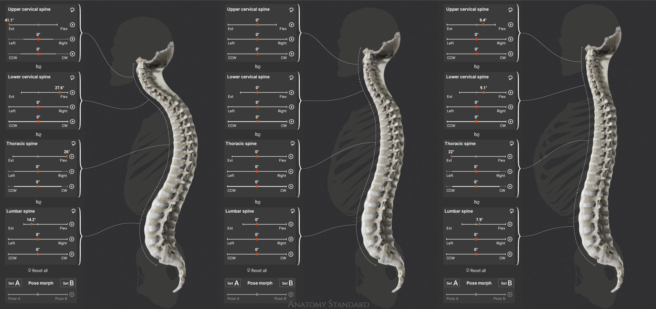
The image above demonstrates the screenshots from the application made with variable adjustments of flexion & extension tuned for different parts of the spine. Choose and click the type of curvature (Straightened, Neutral, or Curved) and see the effect. Note that the resulting stature combines differently directed segmental deformations of the spine within the physiological range. Demonstrated motions occur in the sagittal plane only – this is why other sliders controlling the Left / Right side bending & Clockwise / Counter Clockwise axial rotation remains in place.
Scientific Evidence for the ROM of the Cervical Part of the Spine
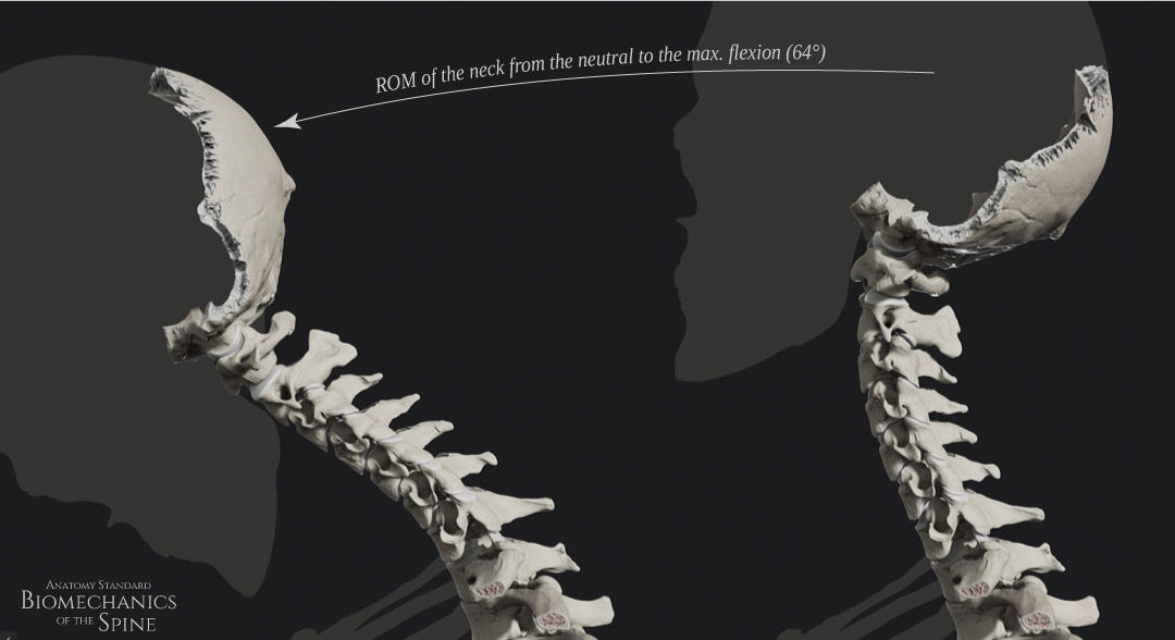
Flexion of the Cervical Spine
The lateral view of the neutral and fully flexed cervical spine (64° of C0-C7 flexion)
Scientific evidence for 64° of cervical flexion based on in-vivo non-invasive studies.
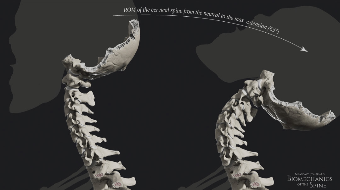
Extension of the Cervical Spine
The lateral view of the neutral and fully extended cervical spine (63° of C0-C7 extension)
Scientific evidence for 63° of cervical extension
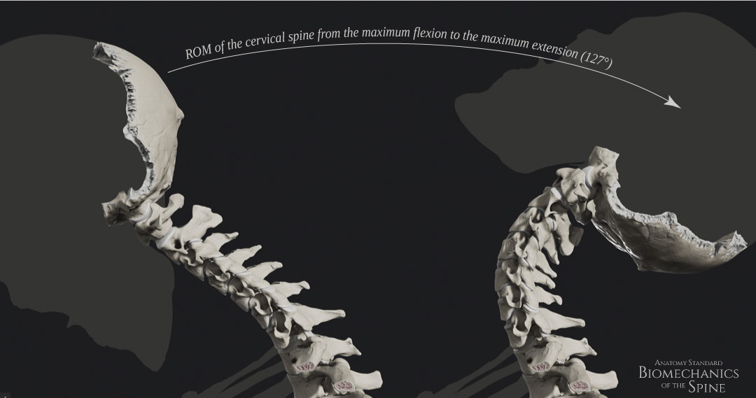
Flexion + Extension of the Cervical Spine
Side-by-side lateral view of the cervical spine motion in sagittal plane – from the full extension to the full flexion (127° of C0-C7 motion)
Scientific evidence for 127° of cervical flexion–extension motion range.
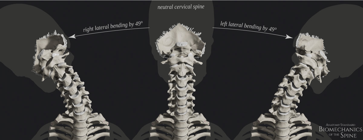
Lateral Bending of the Cervical Spine
Side-by-side anterior view of the cervical spine motion in the frontal plane – from the neutral spine to the right and to the left (C0-C7 one side lateral bending 49°). Note the remarkable axial rotation motion coupled with the lateral bending of the cervical spine.
Scientific evidence for the 49° of cervical lateral flexion motion range.
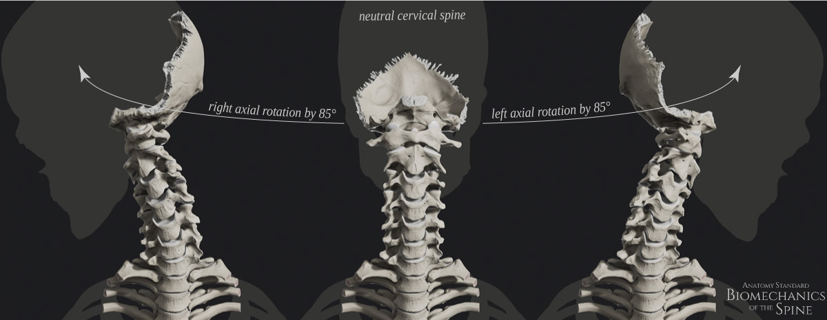
Axial Rotation of the Cervical Spine
Side-by-side anterior view of the cervical spine motion in the horizontal plane – from the neutral spine to the right and to the left (C0-C7 one side axial rotation 85°). Note the substantial lateral bending coupled with the axial rotation of the cervical spine.
Scientific evidence for the 85° of cervical axial rotation range.
ROM of the Thoracic Part of the Spine
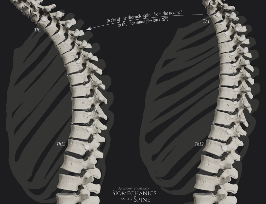
Flexion of the Thoracic Spine
Side-by-side lateral view of the thoracic spine motion in the sagittal plane – from the neutral spine to the full flexion by 26°.
Scientific evidence for the 26° of the thoracic flexion range.
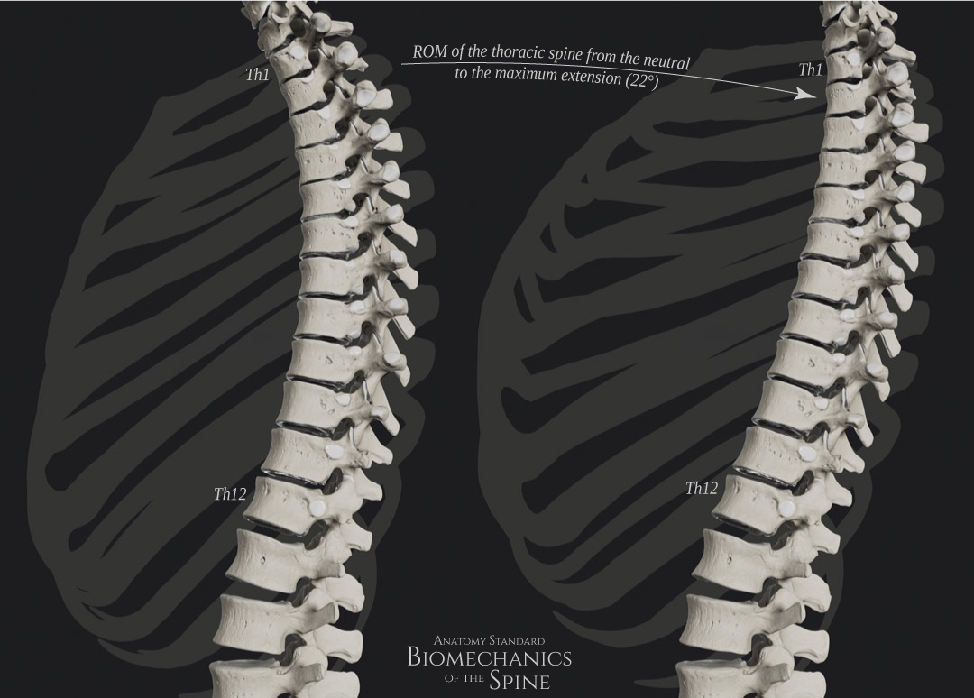
Extension of the Thoracic Spine
Side-by-side lateral view of the neutral and fully extended thoracic spine by 22°.
Scientific evidence for the 22° of the thoracic extension range.
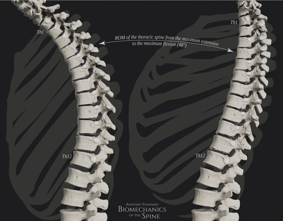
Flexion – Extension ROM of the Thoracic Spine
Side-by-side lateral view of the thoracic spine motion in the sagittal plane – from the neutral spine to the full flexion by 48°.
Scientific evidence for the 48° of the thoracic flexion-extension ROM.
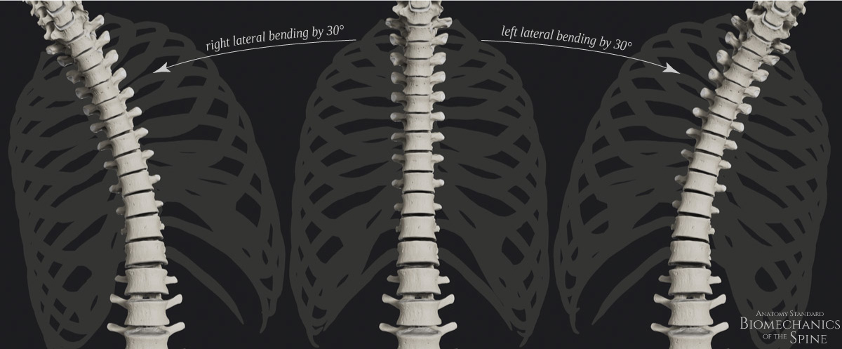
Lateral Bending of the Thoracic Spine
Side-by-side view of the neutral thoracic spine and left / right lateral bending by 30°
Scientific evidence for the 30° of lateral bending of the thoracic spine.
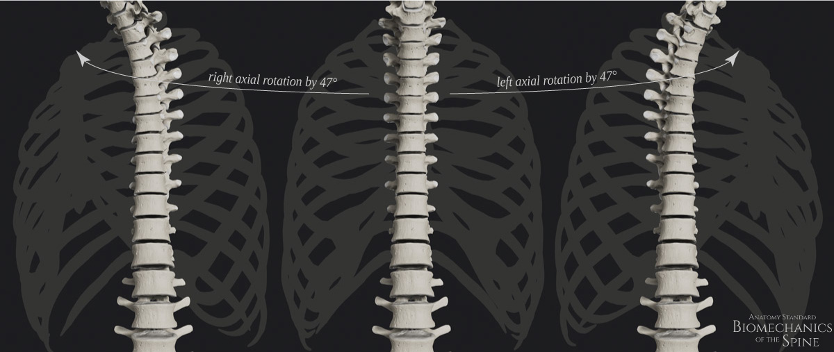
Axial Rotation of the Thoracic Spine
Side-by-side view of the neutral thoracic spine and left / right axial rotation by 47°
Scientific evidence for the 47° of axial rotation of the thoracic spine.
ROM of the Lumbar Part of the Spine
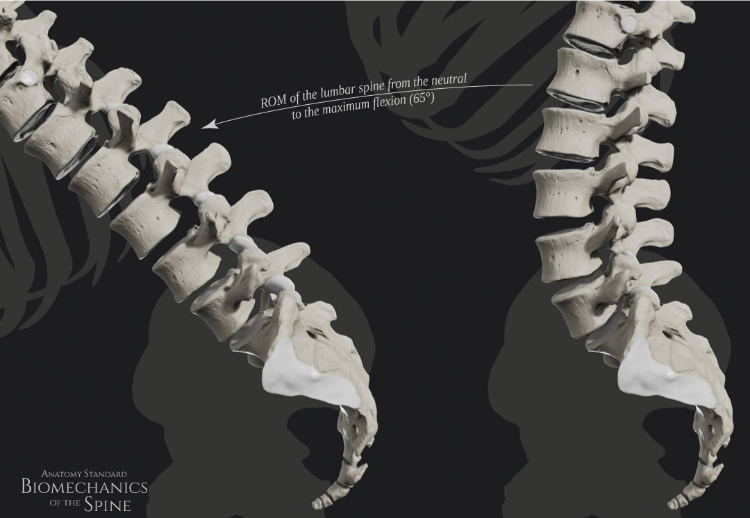
Flexion of the Lumbar Spine
The neutral spine and full flexion of the lumbar spine by 65°.
Scientific evidence for the flexion of lumbar spine for 65°.
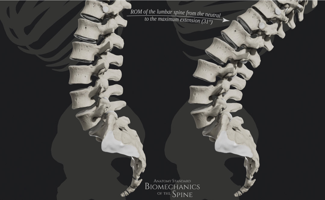
Extension of the Lumbar Spine
Side-by-side lateral view of the lumbar spine motion in the sagittal plane – from the neutral spine to the maximum extension by 31°.
Scientific evidence for the extension of lumbar spine for 31°.
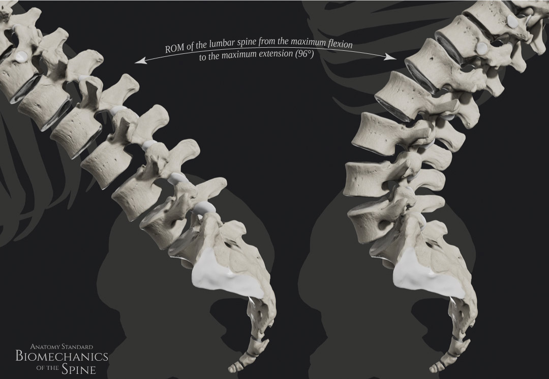
Flexion + Extension of the Lumbar Spine
The ROM of lumbar spine from the full flexion to the full extension by 96°.
Scientific evidence for the flexion + extension range of lumbar spine by 96°.
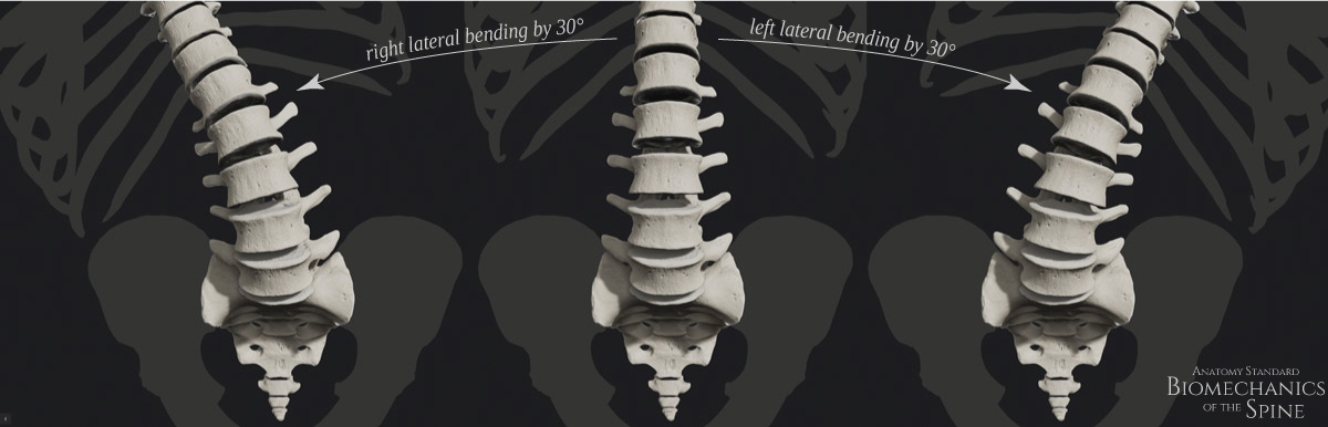
Lateral bending of the Lumbar Spine
Side-by-side view of the neutral lumbar spine and left / right lateral bending by 30°
Scientific evidence for the 30° of lateral bending of the lumbar spine.
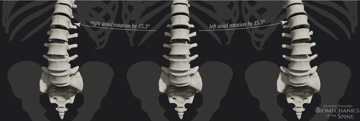
Axial Rotation of the Lumbar Spine
Side-by-side view of the neutral lumbar spine and right / left rotation by 15.3°
Scientific evidence for the axial rotation of the lumbar spine by 15.3°.
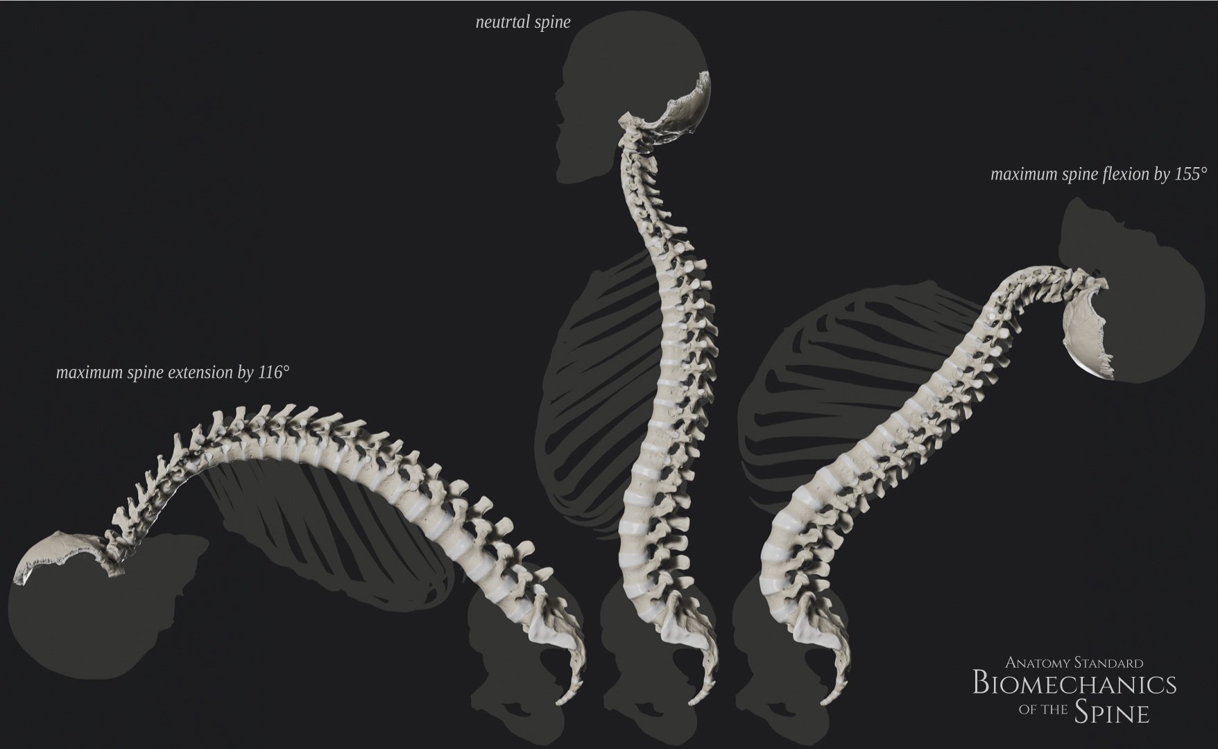
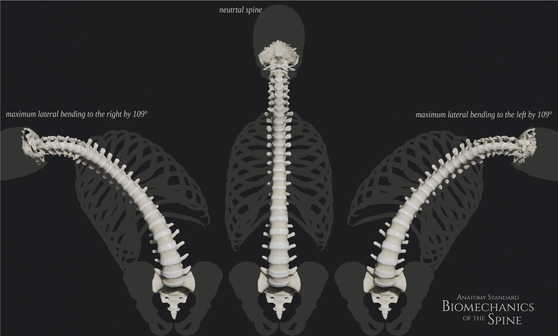
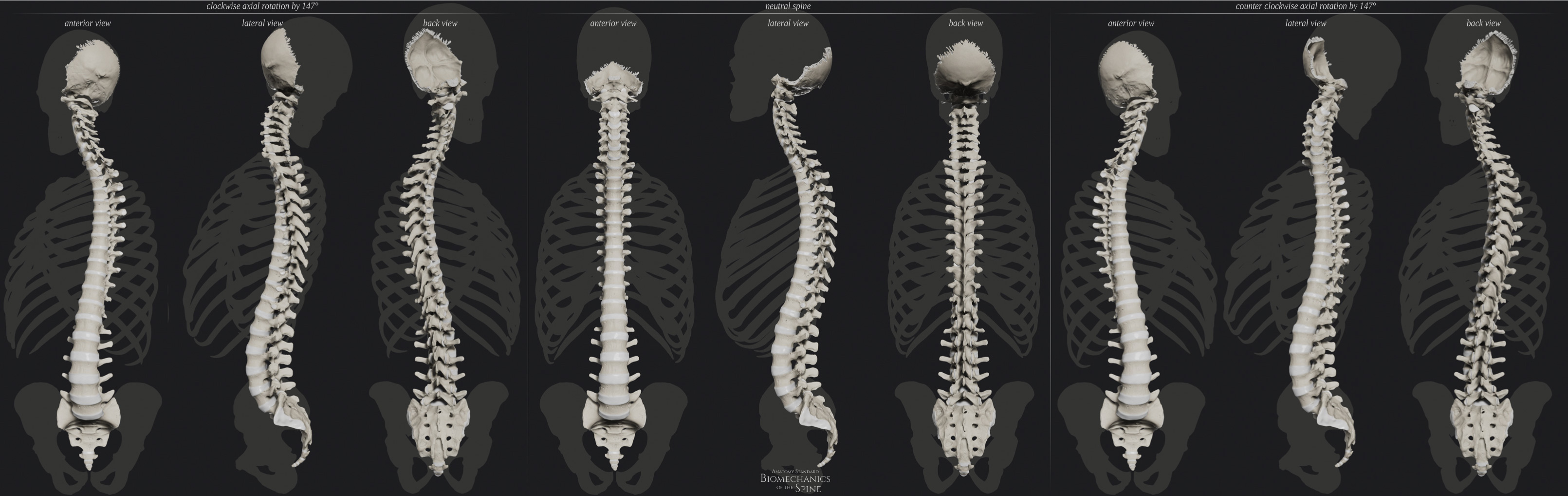
ROM of the Entire Spine in Different Planes
Flexion & Extension
ROM of the spine in the sagittal spine (flexion-extension)
Lewandowski J. Kształtowanie Się Krzywizn Fizjologicznych I Zakresów Ruchomości Odcinkowej Kręgosłupa Człowieka W Wieku 3-25 Lat W Obrazie Elektrogoniometrycznym. Poznan; 2006. ISBN: 8388923633, 9788388923630
_comments.svg)