Centric Occlusion
The term occlusion is not strict, and different authors understand it differently. The simplest definition is limited to "the contact between teeth1." A more precise definition includes static, morphological contact relationship2. A broader definition includes not only the static but also the dinamic (functional) aspects with the perception of occusion as a functional unit, involving teeth, joints and muscles. In the latter case, the concept of occlusion is very closely related to occlusal stability and the homeostatic effect of the maximal intercuspation (centric occlusion)3,4.
The purpose is to demonstrate that the static centric occlusion of our anatomical model is evidence-based and is correctly designed. Centric Occlusion chapter is necessary to fill the gap between the basic overview of the individual teeth and the review of the lateral and protrusive occlusal relation as part of physiological movements of mandible within the functional envelope of motion. These three topics correlate well with the spectrum of occlusion concepts:
| Contact Between the Teeth | > | Static Centric Occlusion | > | Occlusion as an integral part of the Envelope of Mandibular Motion |
Different Aspects of the Centric Occlusion
Dental arches. Overlap of the Teeth.


Click the image to switch between the upper and lower jaws. Both dental arches are shown on the same scale.
The image above demonstrate basic facts about centric occlusion:
First, the dental arches are not uniform in size: the upper jaw is slightly larger than the lower. This results in a marked shift between opposing occlusal surfaces and a reduction in the area of tooth contact (overjet) in a centric occlusion. You can easily check this by touching the edges of molars and premolars with your tongue when the teeth are in full contact – the lower teeth should be closer to the tongue than the upper ones. This overlap is believed to have a protective function: during opening and closing of the jaws, the cheeks, lips and tongue are less susceptible to bite*.
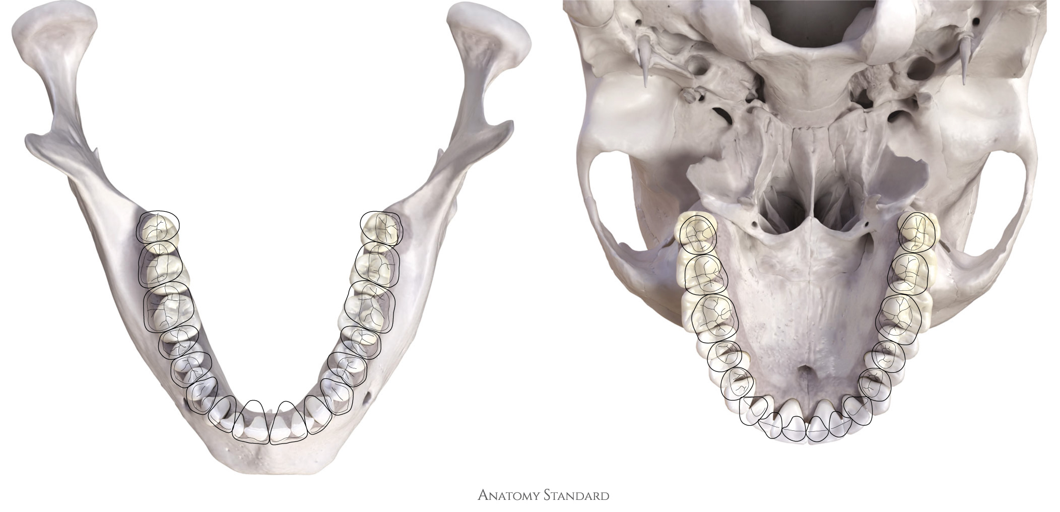
Secondly, the opposed teeth are arranged with an offset in a mesio-distal direction, i.e. the apex of maxillary canine is located between the canine and the 1st premolar of the mandible; the apices of the maxillary 1st premolar are between the mandibular 1st and 2nd premolar etc. This result in the simple fact that (with the exception of the mandibular central incisors and the maxillary third molars) each tooth in one arch contacts two teeth in the opposite arch. This arrangement plays an important role in maintaining the geometry of the arches and in maintaining the occlusal contact with the opposite arch in case of loss, or malposition of tooth*.
Curvatures of occlusal planes.
The occlusal surfaces of teeth do not lie in a plane. Instead, the natural arrangement is intricately curved. Some insight on this is given by the so-called curve of Spee and curve of Wilson:
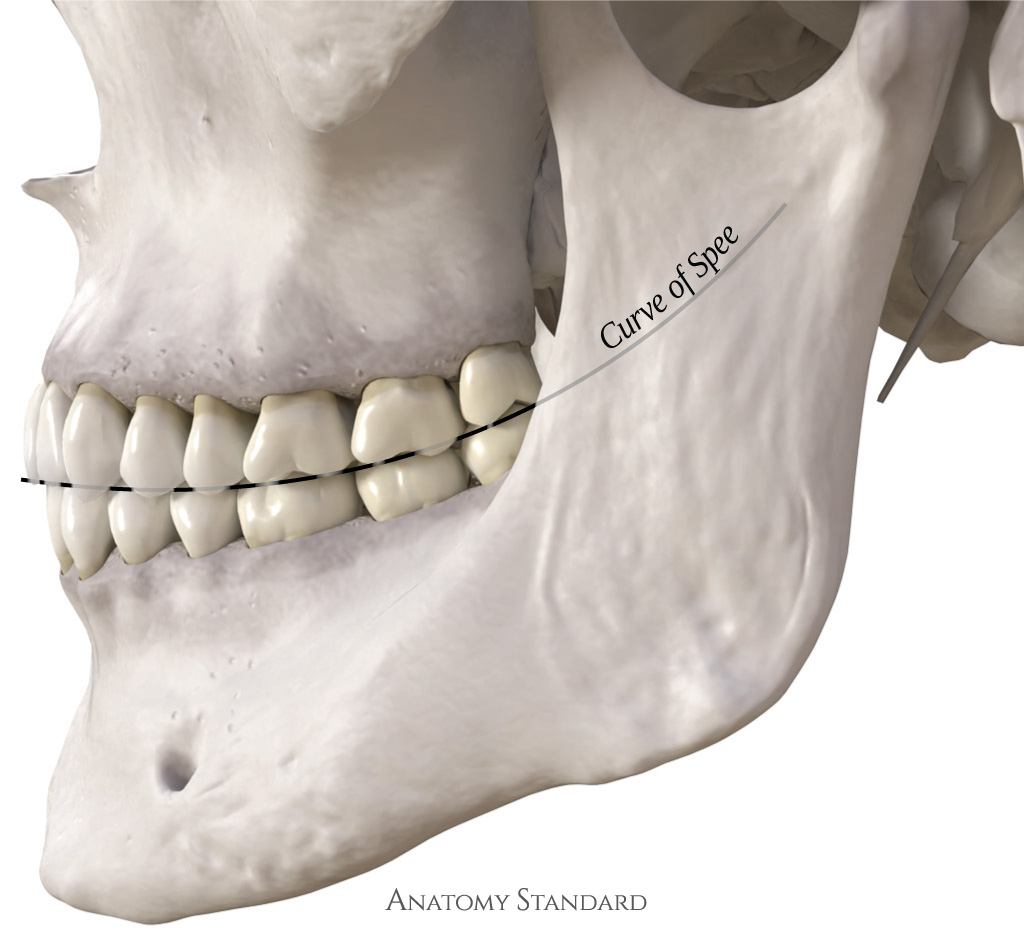

The curve of Spee is a projection of occlusal surfaces onto the sagittal plane. This curve gives the skull a hint of a smile, especially when viewed from the side. The curve of Spee is designed to provide protrusive disocclusion of the posterior teeth through a combination of anterior and condylar guidance*.
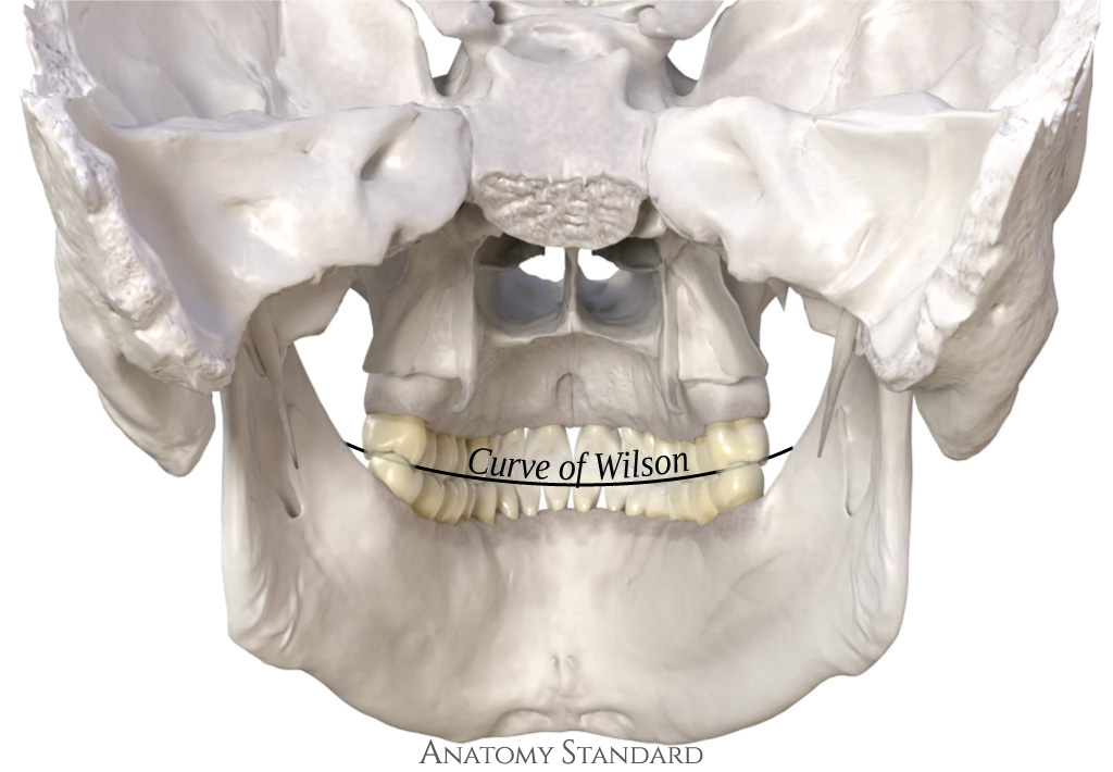

This curve connects the occlusal surfaces of the molars and is observed in the frontal plane*.
The exact degree of curvature of both curves, the curve of Spee and Wilson, varies greatly, and despite attempts to find a universal formula for these curves (Monson spherical theory1 is best known), there is no universal standard that would fit all2,3. There is some limited evidence that the depth of curvature depends on the length of the chewing radius of the mandibular block4,5.
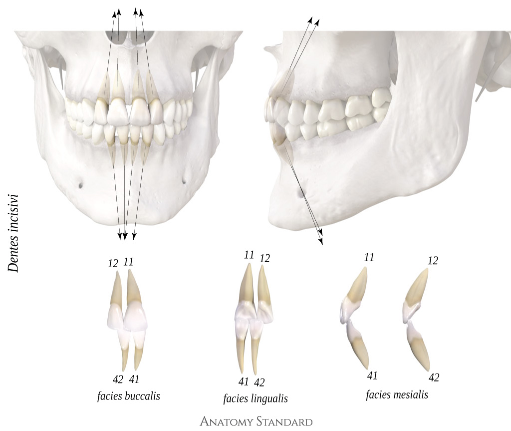
Inclination and angulation of the roots of the teeth
The roots of tooth are not oriented perpendicular to the plane of the occlusive surface. In our 3D model the roots were oriented according with the CT data of a person with the complete permanent denture. Thus, the angulation and inclination of the rooths of our model correlates well with published data*.


Teeth Contacts in Centric Occlusion
In the centric occlusion, the teeth of lower and upper jaws are in contact with each other at several specific points, evenly distributing the occlusal force. These points have been classically described by Dr. Hellman's diagram of 138 points of possible occlusal contacts for a complete set of permanent teeth1. With some modifications, this idealized scheme has been adopted in modern dental anatomy textbooks2 and also implemented in our 3D model of dentition:
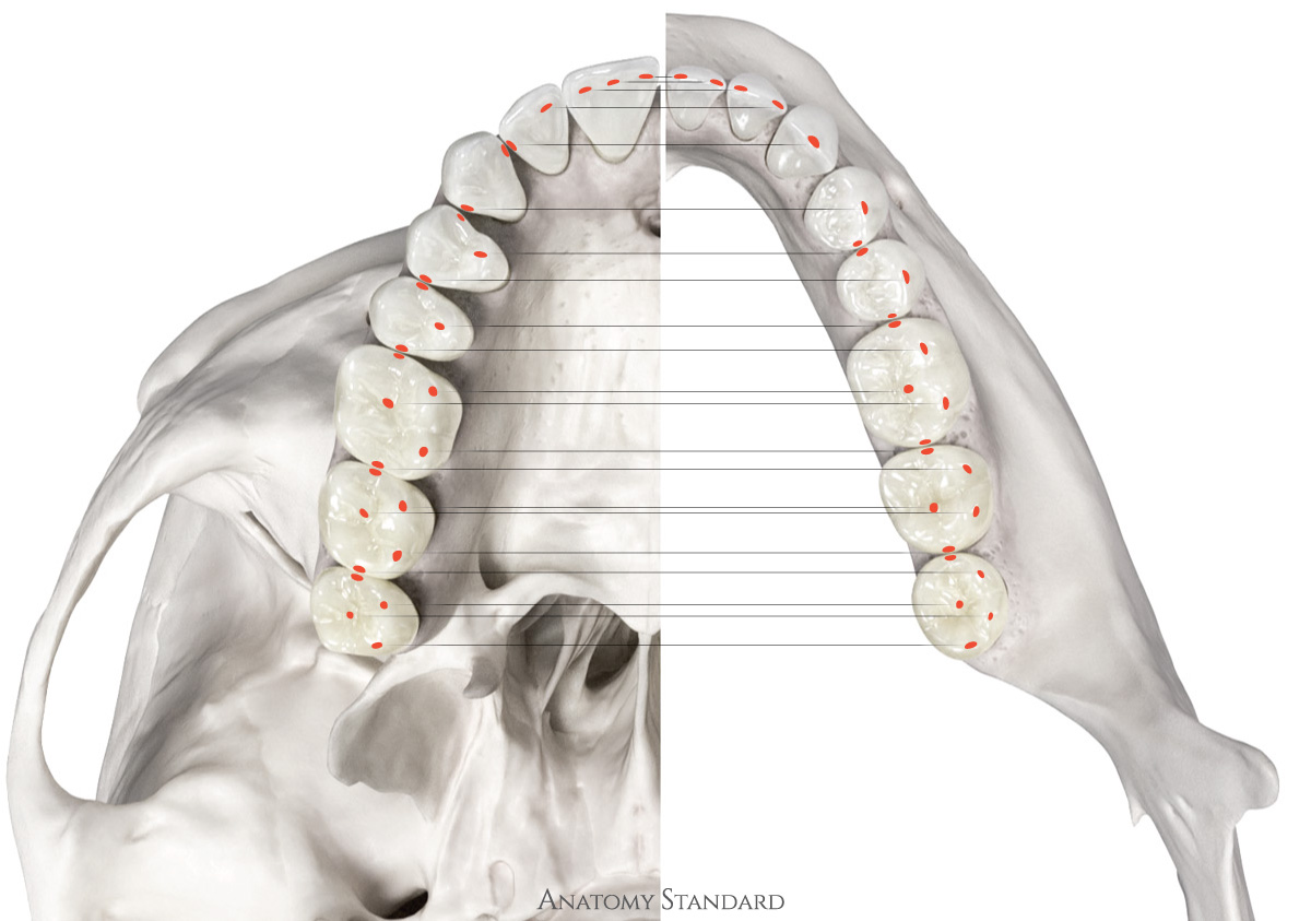

The feature of contact relationship between opposing teeth is that any protrusive or lateral movements of mandible immediately disocclude the premolars and molars. This mechanism is triggered by the incisor / canine guidance in combination with downward translation of the mandibular condyle within the temporo-mandibular joint.