The Bony Pelvis: Anatomy & Sexual Dimorphism
Pelvic skeleton includes two hip bones, sacrum and coccyx.
The geometry of bony pelvis differs significantly between males and females – the phenomena caused by adaptation to the obstetric demands. Females have a relatively larger and rounder pelvic cavity, a shorter and more posteriorly projecting sacrum, a wider subpubic angle, and smaller acetabula with a larger distance between them1,2. It is, however, worth mentioning that in real-life, the sexual dimorphism of the pelvis is not as evident as generally described3,4.
The images presented here demonstrates the classically dimorphic pelvises with the length dimensions based on multiple sources of evidence5–10. The range of reference values reflects rather "average," than "the normal" range, or, by use of statistical terms – rather 95% Confidence Interval, than the double Standard Deviation.

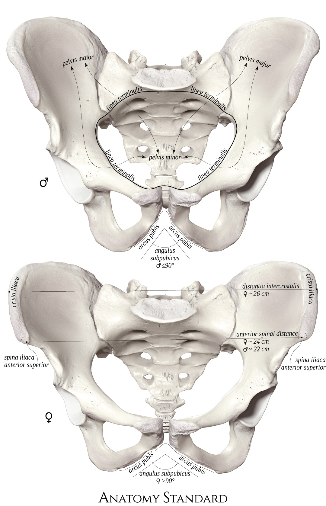


Note the male / female pelvic difference of distance between two spina iliaca anterior superior (anterior spinal distance), the size of the acetabular fossa and the subpubic angle.
The terminal line is a border between the greater and lesser pelvis.
The list of terms:
Pelvis major – Greater pelvisPelvis minor – Lesser pelvis
Linea terminalis – Terminal line
Arcus pubis – Pubic arch
Angulus subpubicus – Subpubic angle
Crista iliaca – Iliac crest
Distantia intercristalis – Intercristal distance
Spina iliaca anterior superior – Anterior superior iliac spine
Anterior spinal distance (iliac bi-spinous diameter)
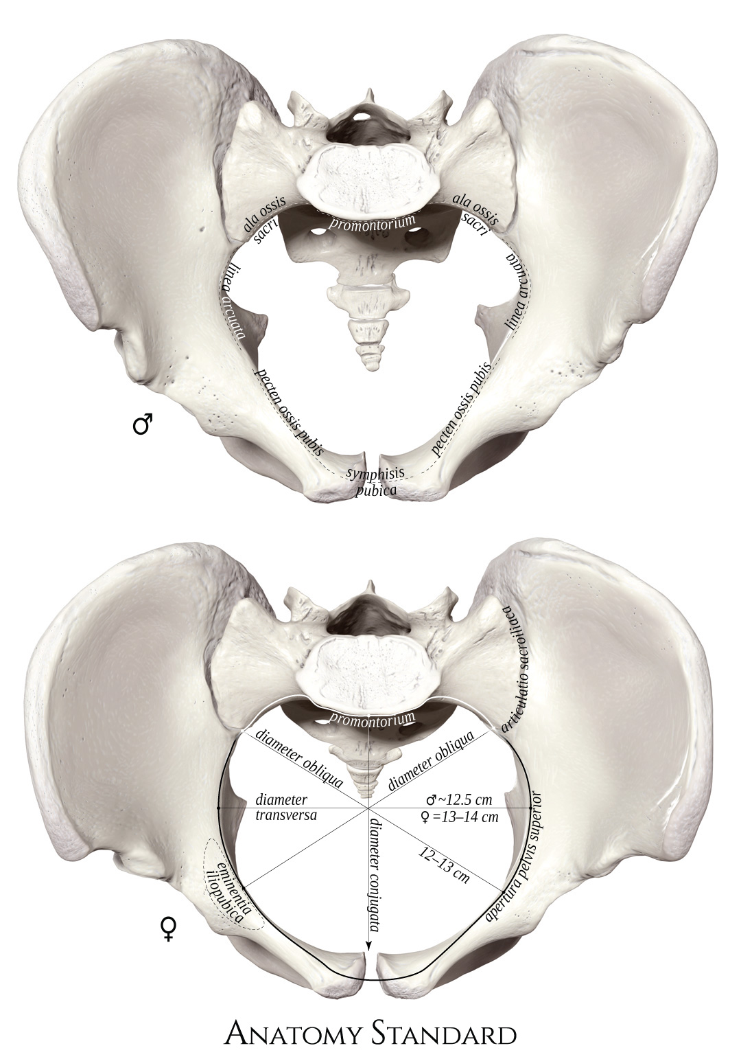


Note the pelvic inlet (apertura pelvis superior) – the upper opening of the lesser pelvis is bordered by the terminal line. The term conjugate apply to the pelvimetric distances in the median plane. The subtypes of conjugates are reviewed below.
The list of terms:
Apertura pelvis superior – Upper opening of lesser pelvis (pelvic inlet)Promontorium – Promontory of sacrum
Ala ossis sacri – Ala sacralis
Articulatio sacroiliaca – Sacroiliac joint
Linea arcuata – Arcuate line
Eminentia iliopubica – Iliopubic (iliopectineal) eminence
Pecten ossis pubis – Pecten (pectineal line) of the pubis
Symphisis pubica – Pubic symphysis
Diameter conjugata – Conjugate diameter
Diameter transversa – Transverse diameter
Diameter obliqua – Oblique diameter
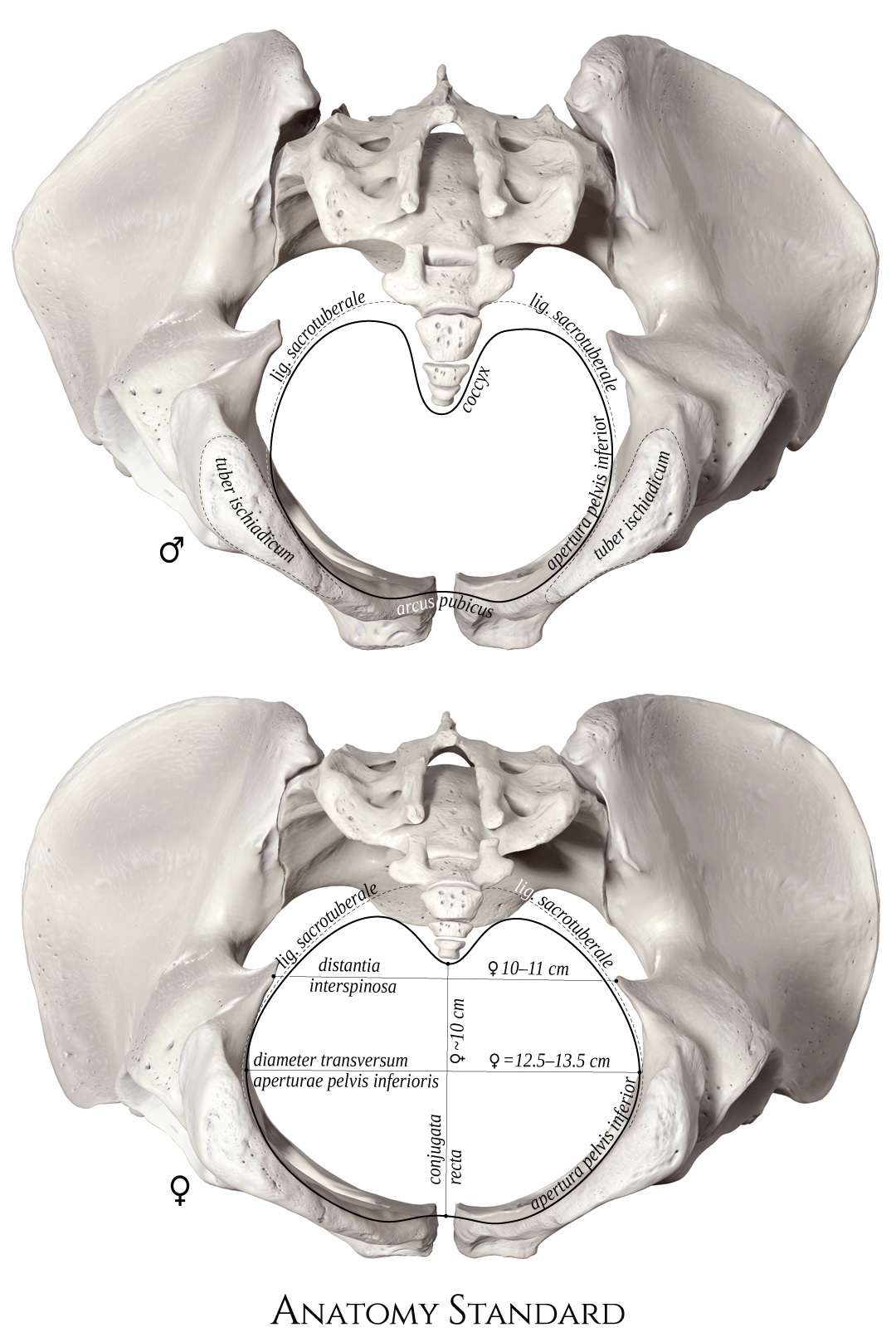


The list of terms:
Apertura pelvis inferior – Lower pelvic aperture (pelvic outlet)Coccyx
Lig. sacrotuberale – Sacrotuberal ligament
Tuber ischiadicum – Ischial tuberosity
Arcus pubicus – Pubic arch
Distantia interspinosa – Interspinous distance
Diameter transversum – Transverse diameter of the pelvic outlet
Conjugata recta – Straight conjugate


Note that anatomical straight conjugate is not particularly relevant. The sacrococcygeal joint is relatively elastic, adding some mobility to the coccygeal bone, so coccyx usually dislocates dorsally during childbirth*.
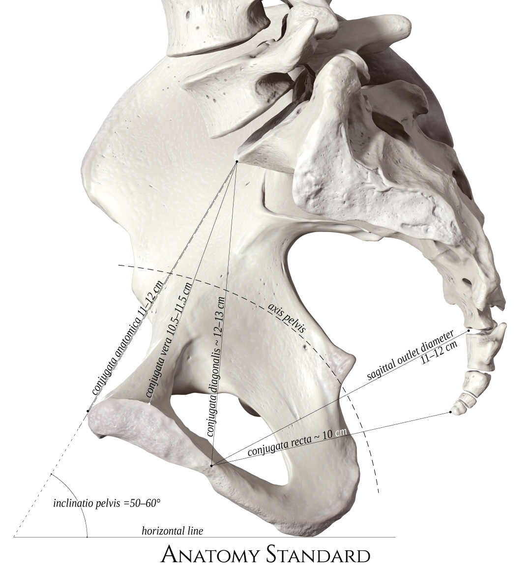
The list of terms:
Axis pelvis – Pelvic axisConjugata anatotmica – Anatomic conjugate
Conjugata vera (gynecologica) – Obstetrical conjugate
Conjugata diagonalis – Diagonal conjugate
Sagittal outlet diameter
Conjugata recta – Straight conjugate
Inclinatio pelvis – Pelvic inclination
Last update: 13/Nov/2025